AAWSAP DIRD, Biosensors and BioMEMS: A Survey of the Present Field, March 2010
DOW-UAP-D136 · page 34 of 45
Text
This text came with the file. It may have mistakes. When the text and the image disagree, the image is right.
UNCLASSIFIED/ /P'OR: OP'P'l@IAL WS& 0Nk¥
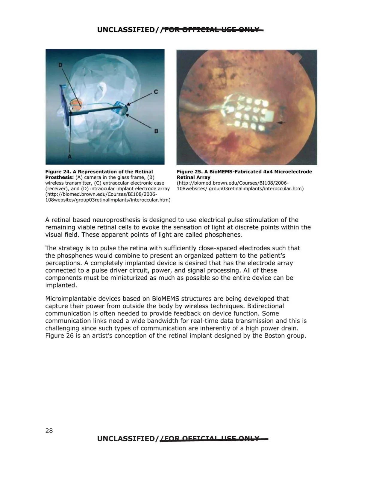
Figure 24. A Representation of the Retinal
Prosthesis: (A) camera in the glass frame, (B)
wireless transmitter, (C) extraocular electronic case
(receiver), and (D) intraocular implant electrode array
(http://biomed .brown .edu/Cou rses/BI108/2006-
108websites/group03retinalimplants/interoccular. htm)
Figure 25. A BioMEMS-Fabricated 4x4 Microelectrode
Retinal Array
(http://biomed.brown.edu/Courses/BI 108/2006-
10Swebsites/ group03retinalimplants/interoccular.htm)
A retinal based neuroprosthesis is designed to use electrical pulse stimulation of the
remaining viable retinal cells to evoke the sensation of light at discrete points within the
visual field. These apparent points of light are called phosphenes.
The strategy is to pulse the retina with sufficiently close-spaced electrodes such that
the phosphenes would combine to present an organized pattern to the patient's
perceptions. A completely implanted device is desired that has the electrode array
connected to a pulse driver circuit, power, and signal processing. All of these
components must be miniaturized as much as possible so the entire device can be
implanted.
Microimplantable devices based on BioMEMS structures are being developed that
capture their power from outside the body by wireless techniques. Bidirectional
communication is often needed to provide feedback on device function. Some
communication links need a wide bandwidth for real-time data transmission and this is
challenging since such types of communication are inherently of a high power drain.
Figure 26 is an artist's conception of the retinal implant designed by the Boston group.
UNCLASSIFIED/ {FOR OFFICIO! !PEii QDlk¥
28