AAWSAP DIRD, Technological Approaches to Controlling External Devices, March 2010
DOW-UAP-D130 · page 27 of 36
Text
This text came with the file. It may have mistakes. When the text and the image disagree, the image is right.
I
I
,
UNCLASSIFIED//P91t 9FFl@IAL YSIE 8HLY
Lamp1 y
bramstem
Amplifiers
Vldoog
A
c=:;)> Recording
,'
I
• .
,
I,
I
,
I
Spike de ecttt>n
rreq
--rt11and low-pass
pulse
fill r
ge
'
omoor
.,_crualC!fs
Additional studies utilizing neurological signals generated from the lamprey have been
equally promising in eliciting cognitive responses that translate a controlled response
into a machine's activity. In a similar experiment, Deliagina, and coworkers (Reference
30) used the activity of reticulospinal neurons recorded from a swimming lamprey to
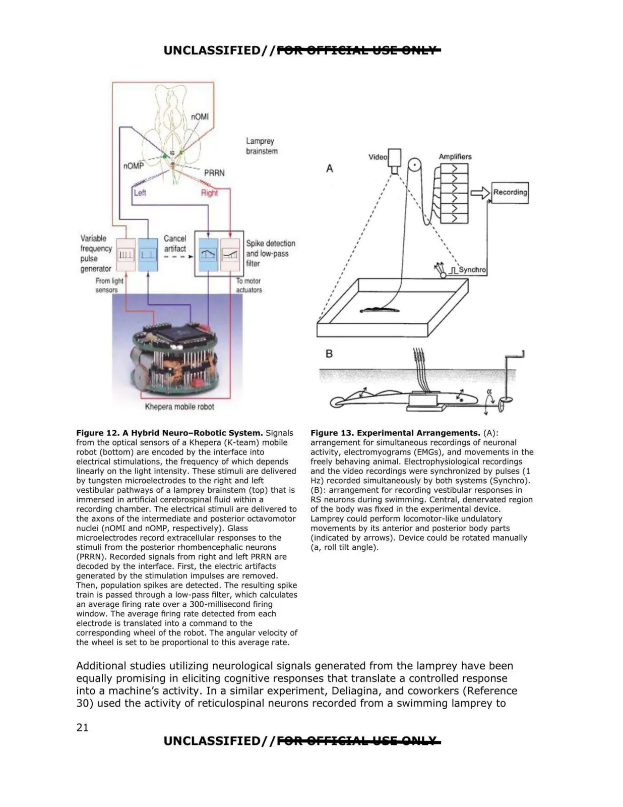
Figure 12. A Hybrid Neuro-Robotic System. Signals
from the optical sensors of a Khepera (K-team) mobile
robot (bottom) are encoded by the interface into
electrical stimulations, the frequency of which depends
linearly on the light intensity. These stimuli are delivered
by tungsten microelectrodes to the right and left
vestibular pathways of a lamprey brainstem (top) that is
immersed in artificial cerebrospinal fluid within a
recording chamber. The electrical stimuli are delivered to
the axons of the intermediate and posterior octavomotor
nuclei (nOMI and nOMP, respectively). Glass
microelectrodes record extracellular responses to the
stimuli from the posterior rhombencephalic neurons
(PRRN). Recorded signals from right and left PRRN are
decoded by the interface. First, the electric artifacts
generated by the stimulation impulses are removed.
Then, population spikes are detected. The resulting spike
train is passed through a low-pass filter, which calculates
an average firing rate over a 300-millisecond firing
window. The average firing rate detected from each
electrode is translated into a command to the
corresponding wheel of the robot. The angular velocity of
the wheel is set to be proportional to this average rate.
Figure 13. Experimental Arrangements. (A) :
arrangement for simultaneous recordings of neuronal
activity, electromyograms (EMGs), and movements in the
freely behaving animal. Electrophysiological recordings
and the video recordings were synchronized by pulses (1
Hz) recorded simultaneously by both systems (Synchro).
(B) : arrangement for recording vestibular responses fn
RS neurons during swimming. Central, denervated region
of the body was fixed in the experimental device.
Lamprey could perform locomotor-like undulatory
movements by its anterior and posterior body parts
(indicated by arrows). Device could be rotated manually
(a, roll tilt angle).
UNCLASSIFIED//F8R. 8FFI&iIAk Uili QNL¥
Variable
ncy
rator
___________
21