AAWSAP DIRD, Biosensors and BioMEMS: A Survey of the Present Field, March 2010
DOW-UAP-D136 · page 37 of 45
Text
This text came with the file. It may have mistakes. When the text and the image disagree, the image is right.
UNCLASSIFIED//P'OR: OP'P'l@IAL WS& 0Nk¥
Variations of pacemaker devices are used in brain neurostimulation for treatment of
Parkinson's disease characterized by the involuntary tremors of the hands and body. In
this case the battery pack must be placed outside the brain, typically in the upper chest
and a long catheter wire tunneled through tissues to the specific parts of the brain to be
treated.
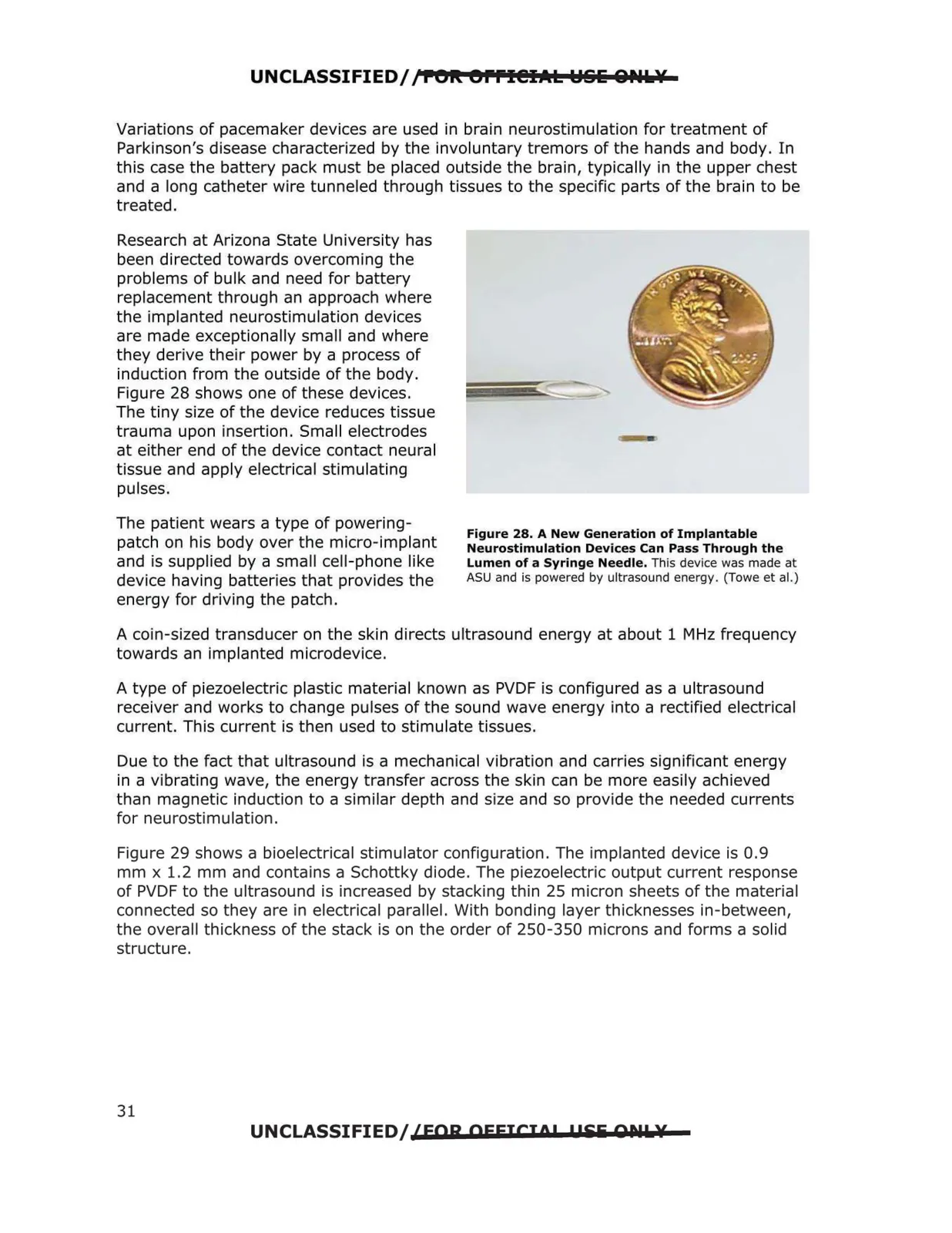
Research at Arizona State University has
been directed towards overcoming the
problems of bulk and need for battery
replacement through an approach where
the implanted neurostimulation devices
are made exceptionally small and where
they derive their power by a process of
induction from the outside of the body.
Figure 28 shows one of these devices.
The tiny size of the device reduces tissue
trauma upon insertion. Small electrodes
at either end of the device contact neural
tissue and apply electrical stimulating
pulses.
The patient wears a type of powering
Figure 28. A New Generation of Implantable
patch on his body over the micro-implant
Neurostimulation Devices Can Pass Through the
and is supplied by a small cell-phone like
Lumen of a Syringe Needle. This device was made at
ASU and is powered by ultrasound energy. (Towe et al.)
device having batteries that provides the
energy for driving the patch.
A coin-sized transducer on the skin directs ultrasound energy at about 1 MHz frequency
towards an implanted microdevice.
A type of piezoelectric plastic material known as PVDF is configured as a ultrasound
receiver and works to change pulses of the sound wave energy into a rectified electrical
current. This current is then used to stimulate tissues.
Due to the fact that ultrasound is a mechanical vibration and carries significant energy
in a vibrating wave, the energy transfer across the skin can be more easily achieved
than magnetic induction to a similar depth and size and so provide the needed currents
for neurostimulation.
Figure 29 shows a bioelectrical stimulator configuration. The implanted device is 0.9
mm x 1.2 mm and contains a Schottky diode. The piezoelectric output current response
of PVDF to the ultrasound is increased by stacking thin 25 micron sheets of the material
connected so they are in electrical parallel . With bonding layer thicknesses in-between,
the overall thickness of the stack is on the order of 250-350 microns and forms a solid
structure.
UNCLASSIFIED/ {FOR OFFICIO! !PEii QDlk¥
31